Mole Mapping: Detect Skin Cancer Early
Mole mapping, also known as total body photography, is a painless, safe, noninvasive approach to help detect melanoma, a dangerous skin cancer. It utilizes photography to record images of the whole body. These images provide dermatologists with a side-by-side comparison of early cancerous moles over time, allowing them to better monitor and track changes such as structure, color and size.
The Importance of Detecting Skin Cancer Early
Changes can be an early sign of melanoma. Early detection is important because melanoma is very treatable in its early stages. When melanoma is detected and removed before it spreads, the cure rate is over 95 percent. Mole mapping also has the potential to reduce the number of unnecessary biopsies for growths that may have slight irregularities that are stable over time and are benign.
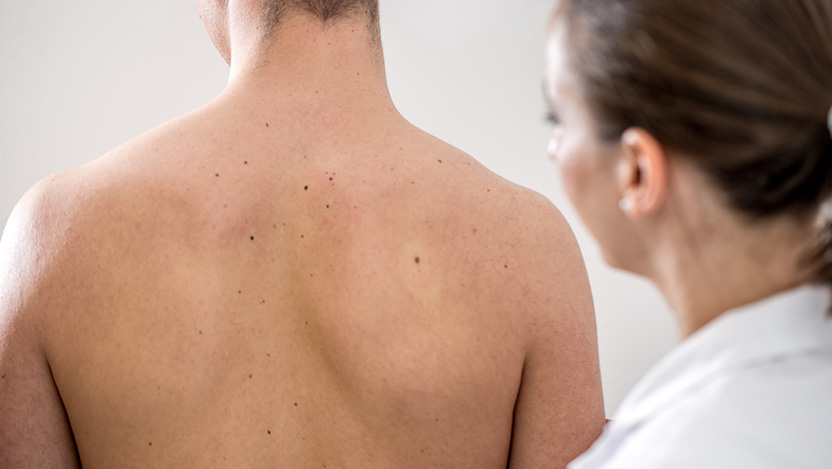
Your First Visit to the Clinic for a Mole Mapping Appointment
A pigmented lesions care team member takes up to 20 digital photos of the patient’s entire skin surface. Then the dermatologist looks for any suspicious moles that should be watched over time. Close-up digital dermatoscopic images of concerning moles are taken at this time. Photographs are stored in a secure database so they can be reviewed for changes at the next visit, which is usually scheduled for 6 to 12 months later.
To prepare for your visit:
- Perform a self-exam and note any new, changing or unusual spots to show the dermatologist.
- Keep in mind the ABCDE’s:
- Asymmetry
- Borders are irregular
- Colors: may have multiple colors
- Diameter greater than ¼ inch
- Evolution of moles
- Discontinue use of any self-tanner/spray tans for one to two weeks before your exam. They may alter the color and pattern of your moles.
- Remove nail polish so the dermatologist can examine the nails on your fingers and toes.
- Wear your hair down so the dermatologist can take a close look at your scalp.
- Do not wear makeup to the visit, or use face wash at the clinic to remove makeup before your exam.
- Do put on moisturizer, which may help normalize the pattern of the moles if your skin is dry.
- Be prepared with questions, such as how to do properly do a skin self-exam.
To inquire about vulvar skin disease or to schedule an appointment, call 773-702-1611.