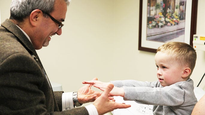
Our expert neurosurgeons created this patient guide on neurovascular lesions to help you better understand your child's condition.
Neurovascular lesions are rare problems that affect the blood vessels in the brain and spinal cord. The most common neurovascular lesions in children are cerebral cavernous malformations (CCM), also called:
- Cavernous angioma
- Cavernous hemangioma
- Cavernoma
A CCM is a formation of fragile, dilated blood vessels that become tangled and form bubble-like structures called caverns. CCM lesions, which are often compared to the shape of a raspberry, may go undetected until they bleed and become symptomatic.
Our team of neurovascular care experts offers an unmatched level specialized care for CCM, including diagnosis, non-surgical and surgical management techniques.
Other pediatric vascular lesions include arteriovenous malformation (AVM) of the brain, which like those in adults, can lead to significant risk of bleeding and potential neurological devastation. The AVM is a tangle of abnormal blood vessels that is prone to bleed or irritate the brain. Symptoms of these lesions include seizures or small hemorrhages.
Once diagnosed, a brain AVM can be treated by surgically removing the vessels feeding the malformations. Before surgery, a radiologist can also place small particles — or "glue" — in the blood vessels that supply the lesions to reduce bleeding during surgery. In addition, children with a small brain AVM may undergo focused radiation treatment or radiosurgery — eventually destroying the abnormal blood vessels and significantly reducing the risk of bleeding.
Learn more about our multidisciplinary vascular anomalies group.
Another infrequent vascular problem in children is moyamoya disease. This syndrome was first seen in children of Japanese descent where the carotid arteries — two of the large feeding vessels to the brain — undergo an unexplained narrowing. Often, this causes stroke or other symptoms of insufficient blood flow to the brain, such as seizures or weakness. The brain responds by developing very small collateral vessels which can appear to resemble a "puff of smoke" on radiographic images. (Hence, the Japanese name moyamoya, which means puff of smoke.)
Usually children with moyamoya respond well to a low-risk surgery called pial synangiosis. During this procedure, neurosurgeons place an artery directly on the brain surface to give the brain additional blood flow. This artery is sewn to the brain and, over time, will develop collateral blood supply to the areas that are in need of additional blood.Request an Appointment
To speak to someone directly, please call 1-773-702-2123. If you have symptoms of an urgent nature, please call your doctor or go to the emergency room immediately.
* Required Field