Breast self-awareness was key to detecting breast cancer

While some may think breast self-examinations are optional, Pam Herber of Yorkville, Illinois, would say that her story is proof that breast self-exams are still an important component of your wellness checklist.
In November 2020, while visiting her son in Oklahoma, 60-year-old Herber reached across her chest and felt something funny. There was a lump in her right breast. With a family history of breast cancer, Herber used to get a mammogram every six months but in the last five years, she would get mammograms yearly and performed self-exams regularly - often enough to immediately recognized something was wrong. When she came back home, she immediately scheduled an appointment with her doctor who sent her for a mammogram.
A mammogram uses low-dose radiation X-rays to detect changes in breast tissue. Because X-rays don’t easily go through breast tissue, the mammography unit uses two plates to flatten the breast to spread the tissue apart. This mechanism creates a better picture and allows radiologists to use less radiation. When symptoms are present, like in Herber’s case, radiologists use a diagnostic mammogram that includes additional images of the breast that are not typically part of a screening mammogram.
Herber’s initial mammogram didn’t show a mass.
Her doctor decided to run a breast ultrasound. Unfortunately, her local hospital didn’t have the breast ultrasound technology available so she had to visit another nearby facility.
A breast ultrasound uses sound waves to draw computer pictures of the inside of the breast. This enables doctors to see any changes in the breast that are difficult to identify with a mammogram. During an ultrasound mammogram, gel is put on the skin of the breast, and a wand, called a transducer, is moved over the skin. The transducer sends out safe sound waves and picks up the echoes as they bounce off body tissues, which are put together to draw a picture of the breast. Breast ultrasounds can tell the difference between fluid-filled cysts, which are usually noncancerous, and solid masses, which may require further testing.
When I told the doctor that I was willing to travel, he told me to go see Dr. Jaskowiak at UChicago Medicine.
Herber’s breast ultrasound showed a mass. The doctors decided to conduct an ultrasound-guided breast biopsy, which uses sound waves to help locate a lump and remove a tissue sample for examination under a microscope.
“It was 2:30 p.m. on New Year’s Eve when my doctor called me and told me that I had breast cancer,” Herber said. “Happy New Year, 2020 sucked.”
Herber’s brother connected her with a doctor that he trusted.
“When I told the doctor that I was willing to travel, he told me to go see Dr. Jaskowiak at UChicago Medicine.”
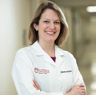
Once Herber established care with Nora Jaskowiak, MD, at UChicago Medicine about 50 miles away from her home, it was like a “well-oiled machine.” With choosing UChicago Medicine for her cancer care, she was able to get another breast ultrasound right at the academic medical center to confirm the findings. She was also connected with plastic and reconstructive surgeon Rebecca Garza, MD Nora Jaskowiak, MD, is an expert in breast surgery and breast cancer, and also specializes in the surgical management of endocrine disorders, including thyroid cancer and parathyroid conditions. She serves as surgical director of UChicago Medicine's Breast Center. Our team represents expertise across the spectrum of breast cancer care: breast imaging, breast surgery, medical and radiation oncology, plastic and reconstructive surgery, lymphedema treatment, clinical genetics, pathology and nursing. Our comprehensive care approach optimizes chances of survival and quality of life. 
Nora Jaskowiak, MD
Breast Cancer Care